Spinal Nerves Spinal Nerve Nerve Spinal Nerves Anatomy Biology Diagrams A 2017 study evaluating the spinal nerve anatomy of 33 deceased people identified spinal nerve plexus variants in 27.3% of them. This suggests that variation is not uncommon and that it doesn't commonly produce noticeable problems. Function . The spinal nerves have small sensory and motor branches. Each of the spinal nerves carries out For the most part, the spinal nerves exit the vertebral canal through the intervertebral foramen below their corresponding vertebra. Therefore, there are 12 pairs of thoracic spinal nerves, 5 pairs of lumbar spinal nerves, 5 pairs of sacral spinal nerves, and a coccygeal nerve. The cervical spinal nerves differ from this pattern. C1-C7 spinal Spinal nerves are bundles of nerve fibers connected to the spinal cord that carry information to and away from the spinal cord. Spinal nerves supply all the areas of the body except most head and neck region (see cranial nerves) with a few exceptions eg neck muscles are supplied by the spinal nerves.. Spinal nerves are mixed nerves sending motor, sensory, and autonomic signals between the CNS

Nerves in Spine Anatomy. The spinal cord serves as a communication highway, linking the brain to the rest of the body through a network of nerve fibers. These fibers branch into pairs of nerve roots that exit through small openings (foramina) between the vertebrae. Each segment of the spinal cord connects to specific regions of the body, which Spinal Nerve Branching. As shown in Figure \(\PageIndex{2}\), axons coming from the posterior (dorsal) root ganglion enter the posterior side through the posterior (dorsal) nerve root.The axons emerging from the anterior side do so through the anterior (ventral) nerve root.The posterior and anterior nerve roots fuse together to form the spinal nerves. Learn about the 31 pairs of spinal nerves that connect the spinal cord to the body's muscles and sensory receptors. Find out how they are named, where they exit from the spine, and what functions they perform.

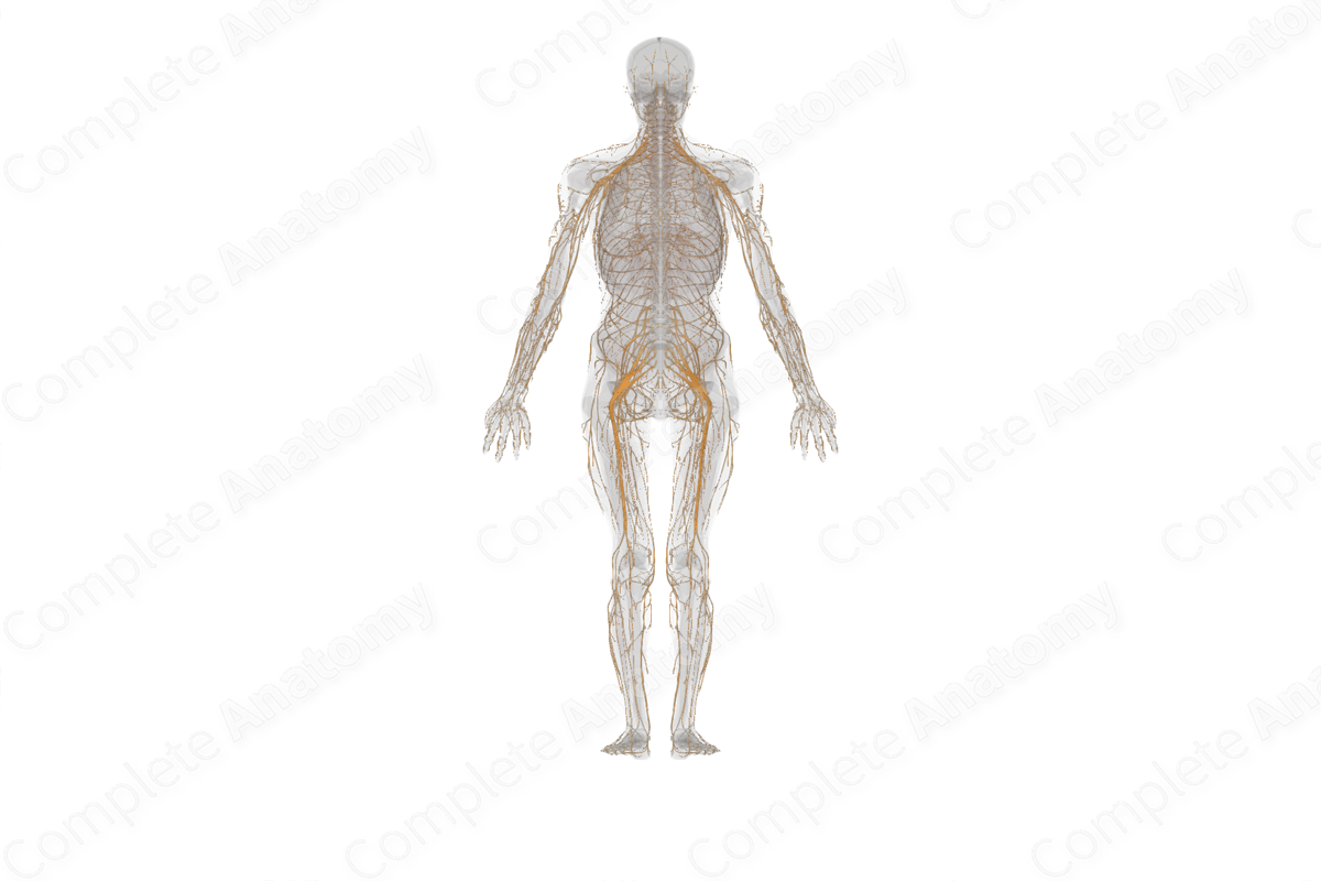
Spinal Nerves Biology Diagrams
Anatomy of Spinal Nerves. Spinal nerves are large nerves that are distributed evenly along the spinal cord and the spine. The spine is a column of vertebrae bones that protects the spinal cord. These spinal nerves are large as they are formed by both sensory and motor nerve roots merging together. These nerve roots emerge from the spinal cord A spinal nerve is a mixed nerve, which carries motor, sensory, and autonomic signals between the spinal cord and the body. In the human body there are 31 pairs of spinal nerves, one on each side of the vertebral column. [1] [2] These are grouped into the corresponding cervical, thoracic, lumbar, sacral and coccygeal regions of the spine. [1]There are eight pairs of cervical nerves, twelve